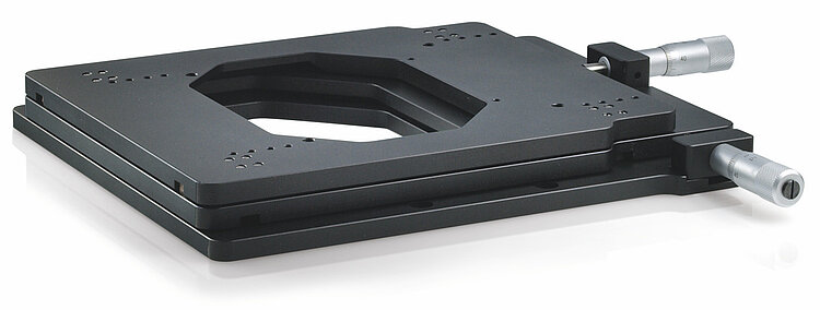
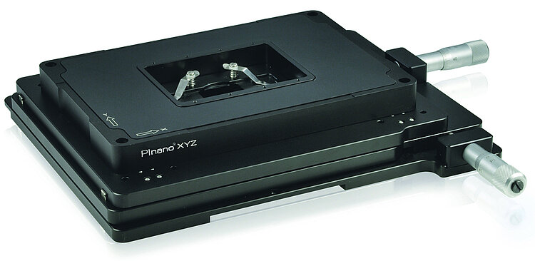
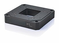
M-545 Microscope XY Stage
Compact, Stable, Long Travel Range
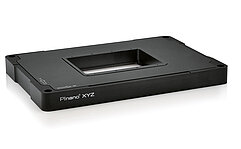
- Stable platform for P-545 PInano® piezo nanopositioning systems
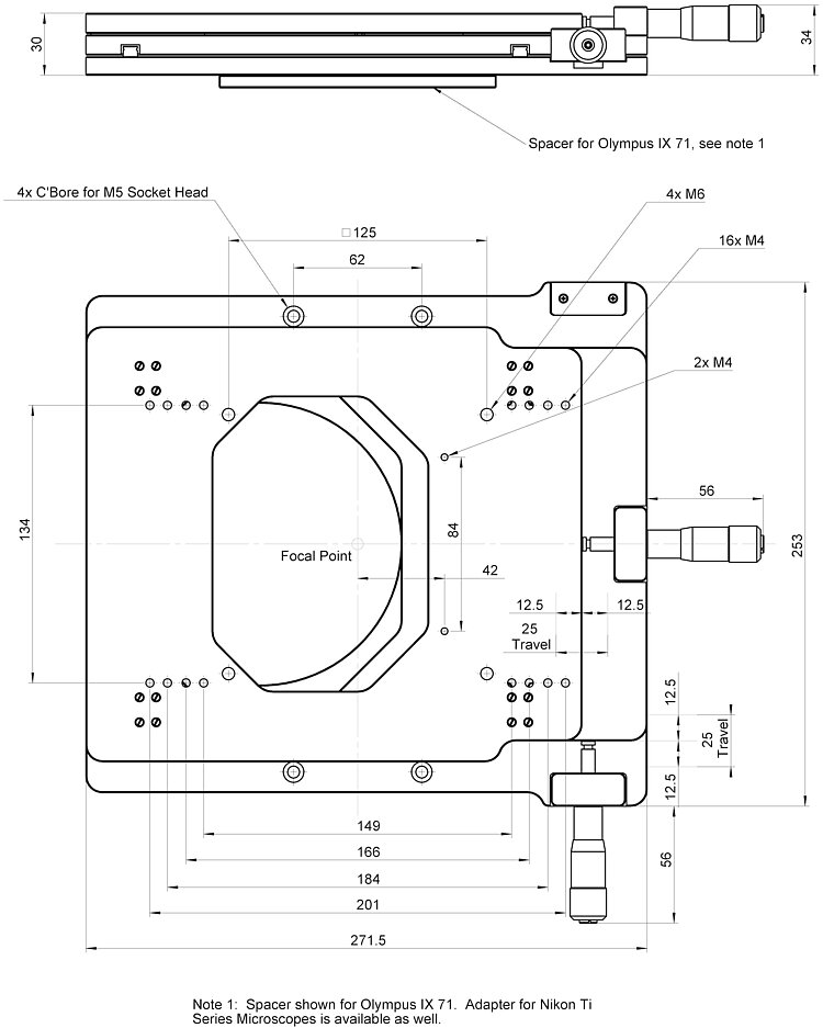
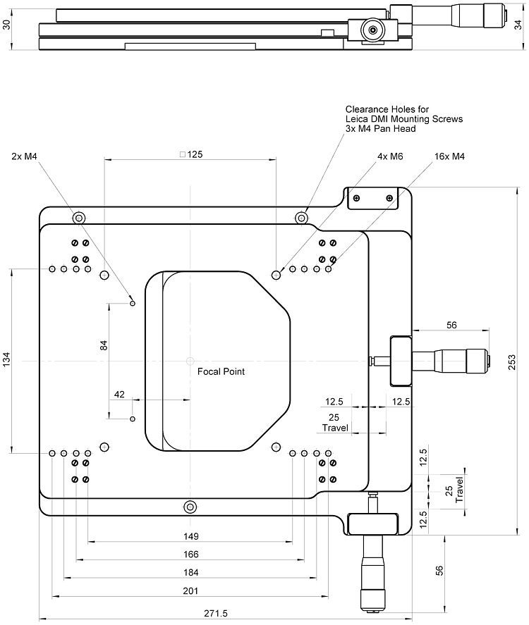
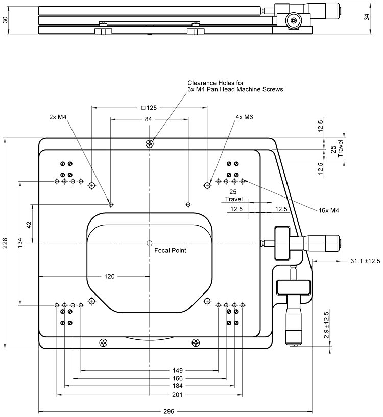
- Low profile for easy integration: 30 mm
- Travel range 25 mm × 25 mm
- Manual operation with micrometer screws, optional motorization
- For inverted microscopes from Nikon, Zeiss, Leica, and Olympus
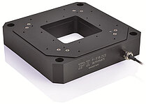
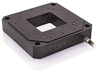
Standard-class, manual microscope XY stage
Rough adjusting for piezo nanopositioners from PI as well as manual positioning of specimen holders. Supports optimum scanning and settling behavior.
Micrometer screw or stepper motor drive
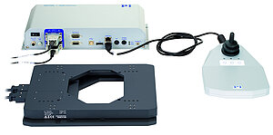
The M-545 XY stage can also be equipped with drives from the M-229 series (all models except M-545.2MZ). Complete packages with two stepper motor drives, matching controller, and joystick, are available under the product numbers M-545.USG respectively M-545.USC.
Suitable for microscopes from a large number of manufacturers
For inverted microscopes from Nikon (TI), Zeiss (Axio Observer), Leica (DMI), and Olympus (IX2, IX3). Versions for other microscopes on request.
Can be combined with numerous PI nanopositioners
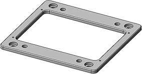
It is possible to mount piezo nanopositioners from the P-545 PInano® and P-541 / P-542 series onto the platform of the M-545. Adapter plates from PI are available for combining with other piezo nanopositioners.
Specifications
Datasheet M-545
Specifications
| M-545.2M | Unit | Tolerance |
|---|---|---|---|
Active axes | X, Y | ||
Motion and positioning | |||
Travel range | 25 mm × 25 mm | ||
Minimum incremental motion | 1 | µm | typ. |
Minimum incremental motion with M-229 linear actuators* | 1 | µm | typ. |
Velocity with M-229 linear actuators* | 1.5 | mm/s | max. |
Mechanical properties | |||
Load capacity | 50 | N | max. |
Preload | 10 | N | |
Miscellaneous | |||
Material | Aluminum, stainless steel | ||
Mass | 4 | kg | ±5 % |
Downloads
Datasheet
Datasheet M-545
Documentation
Installation Instructions M545T0001
M-545.USG / M-545.USC Drive Set for M-545 XY Stage
Short Instructions MP119EK
Positioners with Electric Motors: M-06x, M-11x, M-12x, M-4xx, M-5xx
User Manual PZA02
M-545 Series Microscope Stages
3D Models
M-545 3-D model
Brochure
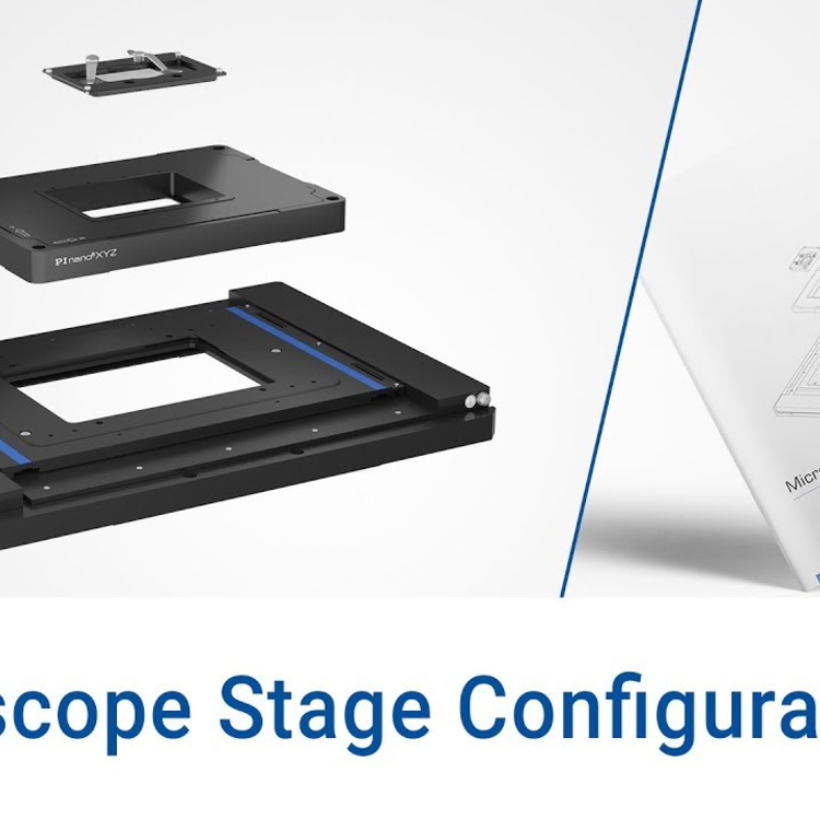
Microscope Stage Configurator
Sample Stages and Holders for Inverted Microscopes
Quote / Order
Ask for a free quote on quantities required, prices, and lead times or describe your desired modification.
Accessories
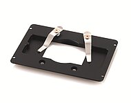
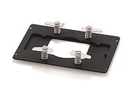
Specimen holders
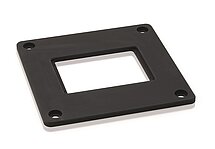
Adapter plates
Stepper motor drive sets (optional; for all models except M-545.2MZ)
Comment obtenir une offre de prix
Contactez un ingénieur!
Quickly receive an answer to your question by email or phone from a local PI sales engineer.
Applications
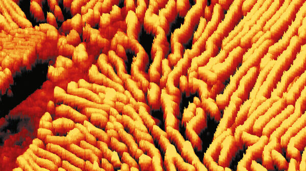
Atomic Force Microscopy
Atomic force microscopy supplies researchers and developers extremely high resolution topographical data from a large number of different minerals, polymers, mixtures, composite materials or biological tissue. This technology, developed in the 1980s, enables users to obtain subatomic resolved images of sample surfaces.

Microscopy & Life Sciences
From genome research through the accurate diagnosis of illnesses to innovative solutions in surgery ‒ life sciences entail numerous disciplines in which new biomedical procedures are researched and devices are developed that are meant to improve the therapy for and quality of life of patients.
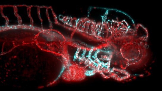
IsoView Light Sheet Microscope
Light Sheet Microscopy is a fascinating technology with a huge application potential in life sciences and biotechnology. IsoView is a brilliant interpretation of this technology, especially intended for imaging fast cellular dynamics across large specimens over several hours. Specimen positioning and objective translation plays a major role in the design of IsoView.
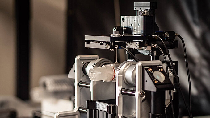
Flamingo Lightsheet Fluorescence Microscopy
Light Sheet Fluorescence Microscopy (LSFM), also called Single Plane Illumination Microscopy (SPIM ) is a very powerful microscopy technology for gentle in vivo imaging offering low phototoxicity and fast image acquisition.
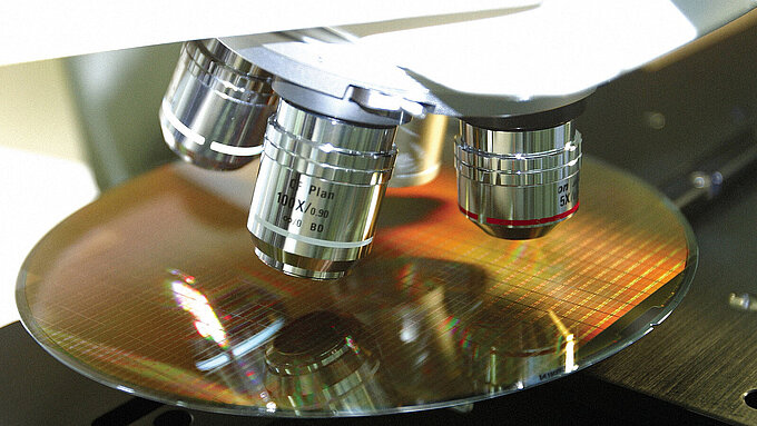
Microscopie à haute vitesse
Le positionnement d'échantillon sur les scanners AFM est réalisé avec des platines piézo dont le rôle est clé pour la qualité des mesures.

GATTAscope (TIRFM)
Les platines linéaires ajustent le faisceau laser dans le microscope TIRF. Un positionnement de précision de l'échantillon est possible en combinant deux platines XY.
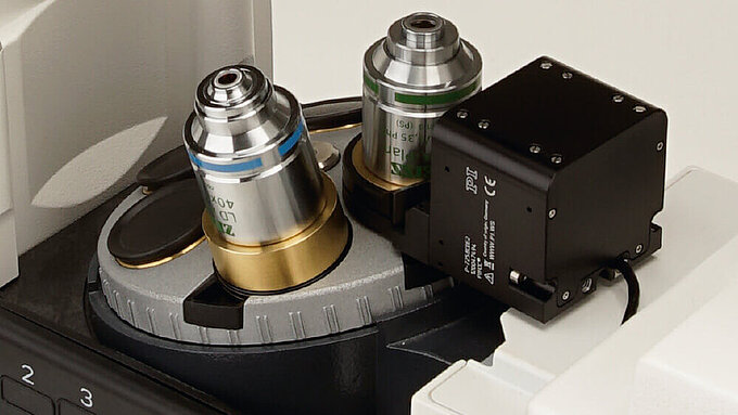
Microscopie confocale
La microscopie confocale est utilisée par exemple en dermatologie afin de détecter la structure de la surface d'un échantillon en déplaçant le plan focal.
